
RISOLNET • KNOWLEDGE ENGINE
SCIENCE & TECHNOLOGY NEWS REVIEW
The November 2 edition of the Risolnet Knowledge Engine explores five developments that demonstrate how scientific progress increasingly depends on the ability to observe, measure and interpret complex systems at different scales.
In astronomy, new observations from the Hubble and James Webb Space Telescopes reveal the same star-forming region through different wavelengths, exposing structures that would otherwise remain hidden. In artificial intelligence, researchers have compared foundation models trained on nearly 1.8 million cellular images, identifying approaches that outperform conventional image-analysis methods in several biological profiling tasks.
Materials scientists have investigated how the mechanical flexibility of perovskite crystals affects the stability of next-generation solar cells. Their findings suggest that the ability of a crystal lattice to accommodate stress may be as important as its electronic properties when designing durable photovoltaic devices.
Meanwhile, research into cryptochrome proteins provides new evidence about the interaction between molecular structure and electron-spin dynamics, offering a possible contribution to the long-standing question of biological magnetoreception.
Finally, a new spatial transcriptomics method has enabled researchers to map approximately 40 million cells across hundreds of mouse-brain sections, demonstrating how large-scale molecular imaging can reveal differences between genetic backgrounds.
The selected developments were originally released or published on October 29 and 30, 2026. This edition distinguishes direct experimental observations, computational predictions, research announcements and potential applications. Scientific findings are not presented as established clinical or commercial technologies unless the available evidence supports that conclusion.
01
Hubble and Webb Reveal the Hidden Structure of the Cat’s Head Nebula
New multiwavelength images of a stellar nursery in the Carina Nebula illustrate how visible and infrared observations reveal different physical processes within the same cloud of gas and dust.

A remarkable cloud of interstellar material within the Carina Nebula has become the subject of a new multiwavelength imaging release from NASA.
The region, informally known as the Cat’s Head Nebula, lies approximately 7,500 light-years from Earth. Its distinctive silhouette resembles a cat viewed from behind, with a narrow pillar of dust forming the body and a bright feature near the upper section resembling an eye.
The images were released on October 29, 2026, and combine observations from the Hubble Space Telescope and the James Webb Space Telescope.
Although the two observatories examined the same region, their instruments recorded different portions of the electromagnetic spectrum. This distinction reveals complementary information about the structure and physical conditions of the cloud.
Hubble’s visible-light observations show a dark, opaque pillar surrounded by luminous gas. The structure appears sharply defined because dense interstellar dust absorbs and scatters much of the visible radiation passing through it.
The surrounding emission includes radiation associated with ionized oxygen, hydrogen and sulfur. Different filters isolate specific wavelength ranges, allowing astronomers to construct a composite image that highlights the distribution of gas and dust.
The pillar is exposed to intense ultraviolet radiation and stellar winds generated by massive stars elsewhere in the Carina Nebula.
These energetic processes gradually erode less dense material through photoevaporation, while denser regions resist destruction and retain their characteristic shapes.
Webb’s Near-Infrared Camera, NIRCam, provides a different perspective. Infrared radiation can penetrate some regions of dust that appear opaque in visible light, revealing additional stars and structural details.
The near-infrared image exposes delicate filaments surrounding the main pillar and shows numerous stars that are difficult to distinguish in the Hubble observations.
Webb’s Mid-Infrared Instrument, MIRI, reveals another aspect of the same environment.
Its observations trace emission associated with warm dust and carbon-containing molecules excited by nearby starlight. The resulting image emphasizes intricate strands, globules and variations in the material surrounding the central pillar.
A bright source near the apparent eye of the cat may be a young star still embedded within the dusty structure.
NASA emphasizes that this interpretation remains provisional. The images alone cannot establish every physical relationship between the bright source and the surrounding filaments.
The importance of the observations extends beyond their unusual appearance.
Star-forming regions are dynamic environments in which gravity, radiation, magnetic fields and stellar winds interact. The resulting structures evolve as new stars form and massive stars reshape the surrounding interstellar medium.
Combining visible and infrared observations allows astronomers to investigate these processes more effectively than either wavelength range could independently.
The Cat’s Head Nebula therefore provides an accessible example of a fundamental principle in observational astronomy: different wavelengths reveal different physical components of the same cosmic environment.
Analysis
Established findings: Hubble and Webb have produced complementary visible-light, near-infrared and mid-infrared images of the Cat’s Head Nebula. The observations reveal differences in dust opacity, stellar visibility and emission associated with illuminated interstellar material.
Interpretation: The morphology of the pillar is consistent with erosion caused by ultraviolet radiation and stellar winds. The bright feature near the apparent eye may be a young star embedded within the cloud.
Limitations: The identification of the bright source as a young star remains provisional. Image morphology alone cannot establish all physical associations between individual structures.
Scientific significance: Multiwavelength observations provide complementary information about the distribution and physical conditions of gas, dust and young stars in active stellar nurseries.
Publication context: Official NASA and STScI imaging release dated October 29, 2026. This is an observational image release, not a newly published peer-reviewed research paper.
02
Self-Supervised AI Models Improve the Analysis of Millions of Cellular Images
A large-scale comparison of computer-vision foundation models identifies approaches that extract biologically meaningful information from microscopy images without relying on conventional cell segmentation and manually designed features.
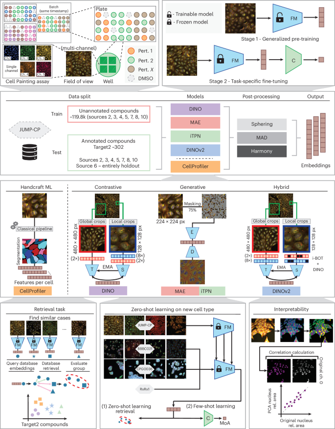
High-content microscopy has become an important tool in pharmaceutical research because it allows scientists to examine how cells respond to chemical compounds and genetic modifications.
One widely used technique is Cell Painting, which combines fluorescent dyes targeting different cellular structures with automated imaging and computational analysis.
The resulting images contain information about nuclei, mitochondria, cytoplasm and other cellular components.
By comparing cellular appearances before and after treatment, researchers can investigate biological activity, identify potentially interesting compounds and explore mechanisms of action.
However, conventional morphological profiling usually requires several computational stages.
Individual cells must first be identified through segmentation. Software then calculates predefined measurements describing their shape, size, texture and fluorescence intensity.
These measurements are subsequently processed to produce numerical representations suitable for statistical analysis and machine learning.
Although effective, this approach can be sensitive to segmentation errors, experimental variability and the limitations of manually selected features.
A study published in Nature Methods on October 29, 2026, evaluates whether self-supervised foundation models can provide a more flexible alternative.
The researchers compared several artificial intelligence architectures, including DINO, DINOv2, masked autoencoders and an integrally pretrained transformer pyramid network.
The models were trained using the JUMP Cell Painting dataset, which includes approximately 1.77 million microscopy images representing 119,876 chemical compounds from seven laboratories.
Unlike conventional supervised learning, self-supervised methods learn useful representations from images without requiring detailed biological labels for every training example.
The researchers then evaluated the pretrained models using independent datasets and compared their performance with CellProfiler, a widely used conventional image-analysis platform.
The evaluation included a held-out collection of 302 annotated compounds and additional datasets covering different cell types, imaging conditions and biological perturbations.
The strongest results came from DINO and DINOv2, which consistently demonstrated better performance than several alternative approaches.
On the EU-OPENSCREEN Bioactives dataset, containing 2,464 compounds, these models achieved improvements of approximately 31 to 37 percent in site-averaged mean average precision for mechanism-of-action retrieval compared with CellProfiler.
The researchers also found that DINOv2 could identify biologically relevant relationships involving compounds targeting proteins such as MAPK14, JAK1 and CDK9 that were not consistently recovered by the conventional baseline.
An important aspect of the investigation concerns generalization.
The models were pretrained on one cellular context but subsequently evaluated using other cell types and experimental conditions.
Their performance suggests that self-supervised representations can capture biological information that extends beyond the original training environment.
Nevertheless, the study also identifies important challenges.
Differences between laboratories, imaging systems and experimental batches can introduce technical variation that interferes with biological interpretation.
The researchers found that postprocessing and batch-effect correction remain important even when sophisticated foundation models are used.
The findings therefore support the use of self-supervised learning as a powerful analytical approach for high-content screening, while emphasizing that reliable biological interpretation still requires careful experimental design and validation.
Analysis
Established findings: The researchers pretrained and compared self-supervised models using approximately 1.77 million images from 119,876 compounds. DINO and DINOv2 outperformed the conventional CellProfiler baseline in several retrieval tasks, including improvements of 31 to 37 percent in a mechanism-of-action benchmark.
Interpretation: Foundation models trained directly on microscopy images can capture biologically relevant cellular representations without requiring conventional segmentation and handcrafted feature extraction.
Limitations: Performance varies across datasets and biological tasks. Differences between laboratories remain challenging, and learned representations can be less interpretable than conventional measurements. The study does not demonstrate autonomous discovery or clinical validation of new drugs.
Technological significance: Self-supervised image analysis may improve the scalability and reproducibility of high-content screening and phenotypic profiling in pharmaceutical research.
Evidence level: Peer-reviewed, open-access research article published in Nature Methods on October 29, 2026. The full research text was available for review.
03
Flexible Perovskite Crystal Lattices Improve the Stability of Tandem Solar Cells
Researchers identify mechanical lattice compliance as a measurable property influencing the resistance of perovskite absorbers to combined heat and light stress, providing a new design principle for bifacial photovoltaic devices.
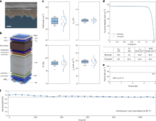
Perovskite-silicon tandem solar cells are among the most promising technologies for improving photovoltaic performance beyond the limitations of conventional single-junction silicon devices.
Their architecture combines two light-absorbing materials with complementary electronic properties.
The perovskite layer captures part of the solar spectrum, while the underlying silicon cell absorbs additional wavelengths that pass through the upper layer.
This arrangement can increase the amount of electrical power generated from a given surface area.
However, the long-term stability of perovskite absorbers remains a major challenge for commercial deployment.
Perovskite materials can undergo structural and electronic changes when exposed to elevated temperatures, illumination and environmental stress.
A study published in Nature Energy on October 29, 2026, investigates a physical property that may help explain differences in stability between perovskite compositions.
The researchers focus on lattice compliance, defined as the ability of a crystal lattice to accommodate mechanical stress through elastic deformation.
Their analysis suggests that perovskite absorbers with greater lattice compliance can better resist certain forms of photothermal degradation.
According to the reported findings, increased compliance reduces the accumulation of thermally induced microstrain within the crystal structure.
It also influences the electronic consequences of structural defects, shifting some deep-level defects toward shallower states and reducing disorder near the electronic band edges.
These effects are important because structural defects can influence charge transport, carrier recombination and the stability of photovoltaic performance.
The researchers applied this design principle to bifacial monolithic perovskite-silicon tandem cells.
Bifacial devices can collect light from both their front and rear surfaces, potentially increasing total energy production when installed above reflective surroundings.
Under a simulated rear-illumination condition corresponding to an albedo-equivalent value of 0.20, the experimental devices achieved a reported power-generation density of 36.2 milliwatts per square centimeter.
This figure represents electrical power per unit area under the specified test conditions. It should not be confused with a power-conversion efficiency of 36.2 percent.
The devices also retained 90 percent of their initial efficiency after more than 1,000 hours of testing under continuous illumination at 65 degrees Celsius.
The results suggest that lattice compliance may provide a useful materials-selection criterion when developing perovskite absorbers designed for demanding operating environments.
Rather than optimizing electronic properties alone, researchers may be able to incorporate mechanical response into the selection and engineering of photovoltaic materials.
However, laboratory stability tests do not establish the operational lifetime of a solar module under real outdoor conditions.
Commercial deployment requires additional evaluation of environmental exposure, encapsulation, manufacturing consistency, mechanical durability and long-term degradation.
Analysis
Established findings: The study identifies a relationship between perovskite lattice compliance and photothermal stability. Experimental bifacial tandem devices achieved a reported power-generation density of 36.2 mW/cm² under specified illumination conditions and retained 90 percent of their initial efficiency after more than 1,000 hours of accelerated testing.
Interpretation: Mechanical flexibility at the crystal-lattice level may help suppress structural degradation and associated electronic losses, offering a measurable parameter for materials optimization.
Limitations: The accessible source was the publisher’s abstract and bibliographic information. Detailed experimental procedures and supplementary datasets were not independently evaluated. Laboratory testing cannot establish commercial module lifetime or field performance.
Technological significance: Integrating mechanical and electronic material properties into photovoltaic design could contribute to more durable perovskite-silicon tandem solar cells.
Evidence level: Peer-reviewed Nature Energy article published October 29, 2026. This report is limited to the information available in the original abstract.
04
Molecular Spin Selectivity Offers a New Clue to Biological Magnetic Sensing
Spectroscopic measurements of cryptochrome proteins reveal a small contribution from chirality-dependent electron-spin polarization, while theoretical calculations suggest that this effect could influence directional sensitivity to magnetic fields.

The ability of migratory animals to orient themselves using Earth’s magnetic field remains one of the most intriguing problems in sensory biology.
Birds and other organisms appear capable of extracting directional information from a magnetic field that is extremely weak compared with many magnetic fields encountered in laboratory experiments.
One proposed explanation involves cryptochromes, a family of proteins that respond to blue light and participate in several biological signaling processes.
Cryptochromes contain flavin adenine dinucleotide, or FAD, which can absorb light and initiate electron-transfer reactions involving nearby amino acid residues.
These reactions can produce pairs of radicals containing unpaired electrons.
The quantum-mechanical spin states of these electrons influence the chemical pathways available to the radical pair.
Because spin dynamics can be affected by magnetic fields, radical-pair chemistry has become a leading candidate for explaining biological magnetoreception.
However, the molecular mechanisms responsible for producing a sufficiently directional and biologically useful signal remain uncertain.
A study published online in The Journal of Physical Chemistry Letters on October 30, 2026, investigates whether chirality-induced spin selectivity could contribute to this process.
Chirality describes the property of a structure that cannot be superimposed on its mirror image. Biological molecules frequently possess this characteristic.
Chirality-induced spin selectivity, commonly abbreviated CISS, refers to the tendency of certain chiral molecular structures to favor the transport of electrons with particular spin orientations.
Researchers led by Yasuhiro Kobori at Kobe University examined a cryptochrome protein from Xenopus laevis using time-resolved electron paramagnetic resonance spectroscopy.
The technique allows scientists to investigate the evolution of electron-spin polarization following light-induced charge separation.
The researchers observed spectroscopic behavior consistent with a measurable contribution from CISS in one of the intermediate charge-separated states.
Their analysis estimated an approximately 8 percent contribution to the observed spin polarization in the secondary charge-separated state.
A smaller contribution, below approximately 6 percent, was associated with a subsequent charge-separated state.
The researchers propose that a subtle twisting rearrangement of an oxidized tryptophan residue contributes to the observed effect.
This interpretation links the physical motion of part of the protein to the quantum-mechanical behavior of its electron-transfer system.
The team also used theoretical calculations to investigate how relatively small spin-selective effects might influence the directional dependence of chemical signaling.
Their model predicted that incorporating a modest CISS contribution could increase the anisotropy of the signaling-state yield from approximately 0.3 percent to 1.6 percent under the investigated high-field conditions.
The calculations therefore suggest a possible amplification mechanism through which small molecular effects could produce more pronounced directional differences in chemical reaction outcomes.
Nevertheless, the distinction between molecular measurements and biological navigation is essential.
The experiments were performed on an isolated cryptochrome system under controlled laboratory conditions, including temperatures substantially below normal physiological conditions.
The theoretical calculations also involved magnetic-field conditions that cannot be directly equated with the Earth’s natural geomagnetic field.
The findings do not demonstrate that living birds use this specific mechanism for navigation.
Instead, they provide molecular evidence supporting the possibility that chirality-dependent spin dynamics contribute to the chemical processes underlying magnetic sensitivity.
Further experiments will be needed to determine whether comparable effects occur under physiological conditions and whether they influence the sensory behavior of living organisms.
Analysis
Established findings: Time-resolved electron paramagnetic resonance measurements support a modest CISS contribution to spin polarization in light-induced charge-separated states of cryptochrome. The estimated contribution reaches approximately 8 percent in the secondary state.
Interpretation: Molecular chirality and conformational motion may influence electron-spin dynamics and contribute to directional sensitivity in radical-pair chemical reactions.
Limitations: The work does not demonstrate magnetic navigation in living animals. Several measurements were performed at low temperatures, and the predicted enhancement in directional response depends on theoretical modeling and specified magnetic-field conditions.
Scientific significance: The findings connect protein conformational dynamics, electron-spin physics and biological signaling, offering a testable hypothesis for future investigations of magnetoreception.
Publication context: Peer-reviewed research published online in The Journal of Physical Chemistry Letters on October 30, 2026. The original research abstract and available scientific text were examined alongside the institutional communication.
05
ATLAS Maps 40 Million Brain Cells to Reveal Differences Between Genetic Backgrounds
A new spatial transcriptomics method combines molecular imaging with computational reconstruction to compare cellular organization across mouse brains, demonstrating a scalable approach to studying tissue architecture and disease models.
Understanding the organization of the brain requires more than identifying which genes are expressed by individual cells.
Researchers must also determine where those cells are located, how different cellular populations are distributed and how their organization changes between biological conditions.
Single-cell RNA sequencing has transformed molecular biology by allowing scientists to characterize gene-expression patterns in individual cells.
However, conventional sequencing methods generally require cells to be separated from their original tissue environment.
As a result, valuable information about spatial organization can be lost.
Spatial transcriptomics addresses this limitation by connecting molecular measurements with the physical location of cells within tissue sections.
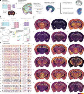
A study published in Nature Methods on October 30, 2026, introduces a method called ATLAS, short for atlas-scale transcriptome localization using aggregate signatures.
The technique combines information derived from single-cell RNA sequencing with in situ molecular measurements and computational reconstruction.
Rather than directly measuring every transcript in every cell, ATLAS uses carefully designed combinations of molecular probes to encode informative transcriptional signatures.
The resulting signals are decoded computationally to infer cellular identities and reconstruct broader transcriptomic information.
This strategy is intended to make large-scale tissue profiling more practical while preserving information about cellular location.
The researchers validated the approach through comparisons with direct measurements obtained using MERFISH, an established multiplexed RNA-imaging technology.
They also compared ATLAS with four additional spatial profiling approaches.
The study then applied the method to a large collection of mouse-brain tissue sections.
Approximately 40 million cells were profiled across more than 400 coronal sections obtained from 15 mice.
The experimental groups included five male and five female C57BL/6J mice, together with five male BTBR mice.
BTBR mice are frequently used as an experimental model for investigating certain behavioral and biological characteristics associated with autism-related research.
The researchers used ATLAS to compare the distribution of cellular populations across the investigated genetic backgrounds and biological groups.
Their analysis identified more than 40 statistically significant differences in cell-type distributions.
They also reported 16 regional changes in cellular composition across the investigated comparisons.
These findings demonstrate the ability of the method to detect differences in tissue organization that may be difficult to characterize using approaches lacking comparable spatial coverage.
An important feature of ATLAS is the use of computational inference.
The method relies on reference transcriptomic information to reconstruct cellular identities and estimate molecular features from a compressed set of measurements.
Consequently, its results depend partly on the quality and biological relevance of the reference datasets and decoding procedures.
The technique should not be interpreted as directly measuring every gene transcript in each of the 40 million cells.
Nevertheless, the study demonstrates how molecular imaging, computational analysis and reference atlases can be combined to investigate tissue organization at substantial scale.
Potential future applications include comparative studies of developmental biology, genetic variation and experimental disease models.
Additional validation will be required before the method can be extended confidently to other tissues, species or clinical research environments.
Analysis
Established findings: ATLAS was used to profile approximately 40 million cells across more than 400 mouse-brain sections. The study identified more than 40 significant differences in cell-type distributions and 16 regional composition changes across the investigated comparisons.
Interpretation: Combining spatial molecular measurements with computational reconstruction can support large-scale comparisons of cellular organization across genetic backgrounds.
Limitations: The accessible publisher content consisted primarily of the abstract, figure descriptions and bibliographic information. ATLAS uses computational inference rather than direct measurement of every transcript. The reported biological findings concern mouse models and cannot be directly generalized to human neurological conditions.
Life-science significance: Scalable spatial transcriptomics could help researchers investigate how genetic variation influences the organization of complex tissues and biological systems.
Publication context: Peer-reviewed Nature Methods article published October 30, 2026. Quantitative claims in this report are limited to the original abstract and accessible publisher information.
The RKE Signal
The central theme of today’s edition is the growing importance of extracting reliable information from systems that are too complex to understand through direct observation alone.
The Cat’s Head Nebula illustrates this principle at astronomical scales. Visible-light observations reveal the external structure of a dense dust cloud, while infrared measurements expose stars and material that would otherwise remain hidden. Neither observation is complete by itself. Together, they provide a more informative representation of the same physical environment.
The artificial intelligence study demonstrates a related transformation in biological image analysis. Instead of relying exclusively on predefined measurements, self-supervised models learn representations from large collections of cellular images. These representations can reveal relationships between chemical perturbations that are difficult to capture through conventional feature engineering.
However, the results also show that sophisticated computational models do not eliminate experimental variability. Differences between laboratories and imaging conditions remain important sources of uncertainty.
The photovoltaic research extends the discussion from information processing to materials engineering. The mechanical response of a crystal lattice influences the stability of its electronic behavior. A material’s performance cannot therefore be understood exclusively through its electrical characteristics.
This relationship is particularly relevant to emerging energy technologies, where laboratory efficiency must eventually be reconciled with durability, manufacturing consistency and real operating conditions.
The cryptochrome investigation provides another example of interactions across physical scales. Molecular motion may influence electron-spin polarization, while spin-dependent chemical reactions could contribute to the generation of directional biological signals.
The study does not solve the mystery of animal magnetoreception, but it identifies a molecular mechanism that can be investigated experimentally.
Finally, ATLAS demonstrates the value of combining molecular measurements with computational reconstruction. By mapping tens of millions of cells while preserving their spatial organization, researchers can investigate tissue-level differences that would be difficult to interpret from isolated cellular measurements.
The method also illustrates an important distinction between direct measurement and inference. Reconstructing molecular information from compressed measurements can provide substantial analytical advantages, but the accuracy of the resulting maps depends on reference data, experimental quality and computational assumptions.
Across these five developments, a common scientific pattern emerges: progress often comes from combining complementary forms of evidence rather than searching for a single definitive measurement.
Astronomical imaging, artificial intelligence, materials characterization, molecular spectroscopy and spatial transcriptomics all depend on carefully interpreting signals produced by complex physical or biological processes.
The RKE Signal for November 2, 2026: the next generation of scientific discovery will depend not only on collecting more data, but on understanding how those data were generated, what they genuinely measure and where the boundaries of interpretation remain.